

LVivo AI Toolbox™
Triage and Monitor COVID-19 Patients' heart
How COVID-19 Impacts the Heart
Clinical protocols from the American and European Societies of Echocardiography recommend conducting focused echo exams to assess heart function in patients on the emergency COVID-19 frontlines.
The Challenge: Cardiac Ultrasound Analysis During COVID-19
Point of care ultrasound (POCUS) is helping clinicians identify and monitor COVID-19 patients' heart symptoms on the frontlines.
Clinicians need automated solutions to support decisions at COVID-19 patients' bedsides.

Variability
Ultrasound analysis at Point-of-care is still mostly done visually, in a subjective process that is both prone to error and highly dependent on the user's experience

Bottlenecks
The rising number of cardiac procedures needed is creating a load on all clinicians, while clinical guides recommend minimizing scans and contact with patients

Recent data show that 51% of hospitalized COVID-19 patients who died had heart damage

Clinicians believe what causes heart problems identified in COVID-19 patients to be one of the pandemic's “critical unknowns"
Clinical protocols recommend performing limited cardiac ultrasound scans including Left and Right ventricles analysis to detect cardiac dysfunction in patients with suspected or confirmed COVID-19.
Our FDA Cleared and CE Marked solutions, LVivo EF™ and LVivo RV™, provide automated, quantified analysis of the Left and Right Ventricle, including key measurements about each ventricle’s size and function.
Case study: DiA on the frontlines
A 66 y/o COVID-19 patient arrived at the ED with tachycardia and an ECG showing T-wave abnormality.
LVivo EF on Vscan Extend ultrasound delivered immediate results of normal Ejection Fraction (55%) which supported the physician's conclusion that viral myocarditis as the possible cause was unlikely.
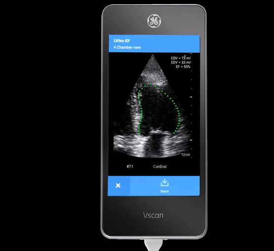
LVivo EF
Ejection Fraction is a key indicator of left ventricular cardiac dysfunction, which protocols recommend monitoring to help triage COVID-19 patients.
LVivo EF by DiA provides automated analysis of the left ventricle, from 4CH and/or 2CH views, including single and Biplane: EF, End-Systolic Volume (ESV), End-Diastolic Volume (ED) Stroke Volume and Global Longitudinal Strain.


LVivo RV
Studies show that assessing the heart’s RV size and function is a key indicator of cardiac dysfunction including pulmonary embolism and heart failure common in COVID-19 patients.
LVivo RV provides automated analysis of the right ventricle, from 4CH modified/ focused apical view including: Fractional Area Change (FAC), EDA, ESA, Free Wall Strain and TAPSE.

Objective and reproducible analysis of cardiac function
Faster and more confident clinical decisions - conserve additional resources needed

Reduce scan time, load on medical staff and bottlenecks
Minimize patient contact and potential exposure
to infection

Ziv Dadon, M.D
Department of Cardiology
“COVID-19 has in many ways made clinicians more open to embracing new technologies that can help them work better and smarter...AI makes an important difference and I think a solution like LVivo EF has a central place in ultrasound analysis of the left ventricle.”
“COVID-19 was a great pilot in terms of increasing awareness of how AI can assist with diagnosis. Being able to assess cardiac contractility Ejection Fraction rapidly is really important. An AI-driven tool like LVivo is going to improve the ability to rapidly triage and evaluate patients.”

Bret Nelson, M.D
Department of Emergency Medicine

“LVivo EF is a great app. I have used it a lot on ICU during COVID-19 to highlight some of the effects of the virus on the cardiac muscle.”
Jonny Wilkinson, M.D
Intensive care and anaesthesia

"LVivo EF makes it possible for clinicians to quickly, accurately and safely analyze cardiac function at bedside. For patients with COVID-19...being able to rapidly and accurately detect the EF at the point of care can save precious time and improve patient outcomes."
Evan (Avi) Alpert, M.D
Department of Emergency Medicine
Our Partners
DiA's AI solutions run on the CPU and GPU of any ultrasound device or Healthcare IT system (PACS/ Cloud).


COVID-19 Resources
Partner with DiA to realize our COVID-19 mission
The time for AI-powered ultrasound is now. We’re proud to be working with
our partners and clinicians across the globe to maximize the benefits of AI
in the fight against COVID-19.
If you are an ultrasound user, vendor, or PACS company, please join us to help clinicians and patients on the COVID-19 frontlines.